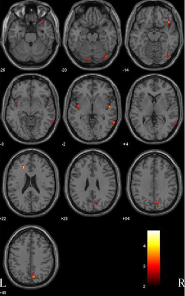
Figure 3.

Gray matter comparison between patients with Parkinson disease with mild cognitive impairment (PD-MCI) and normal controls (NC). Significant changes are found in the fusiform gyrus and lingual gyrus (both sides), anterior cingulate and insular cortex (left side), and right superior temporal gyrus, orbitofrontal cortex, central gyrus and precuneus (right side). L, left side; R, right side. The color bar represents the T score. Yellow presents a high T score.
