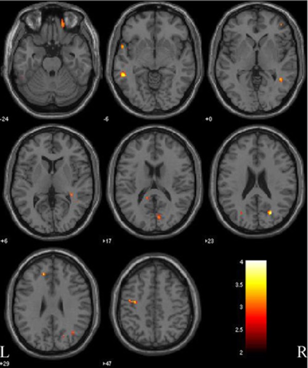
Figure 4.

Gray matter comparison between patients with Parkinson disease with and without mild cognitive impairment (PD-MCI versus PD-nMCI). Significant changes are observed in precentral gyrus and middle temporal gyrus (both side), cuneus, precuneus, and orbitofrontal cortex (right side), and fusiform gyrus (left side). L, left side; R, right side. The color bar represents the T score. Yellow presents a high T score.
