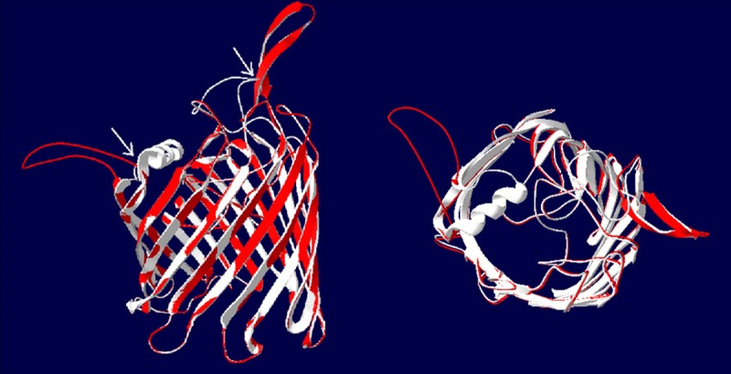
Figure 3.

The three-dimensional structure of OprD. The PCR products of OprD genes from 29 MDR-AB strains were sequenced, and the homology analysis showed that there were mutations in MDR-AB strains compared with the SDF strain. It was significant difference in three dimensional structure of protein between MDR-AB and SDF strains by molecular modeling and comparison of overlapping molecular structure. The white region isthe structure of OprD and the red region is the structure of MDR-AB. Left: the frontage of β-barrel structure; Right: the side of β-barrel structure.
