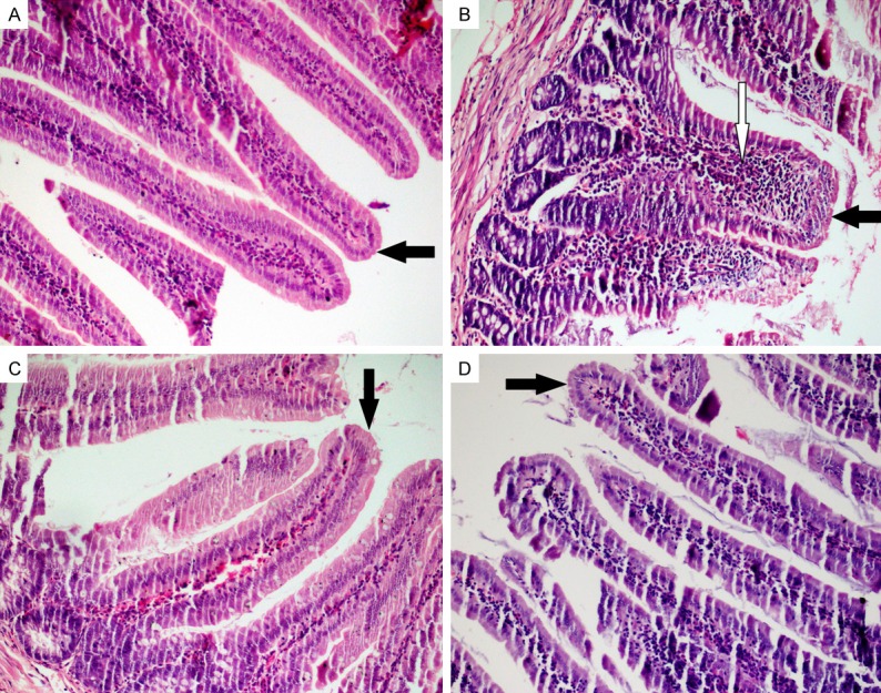
Figure 1.

Staining with hematoxilin eosin (HE). A. Control; sharp-edged and long villi, normal cellularity with lamina propria; B. MTX; shortening and fusion of the villi, crypt loss and significant inflammatory cell infiltration in the lamina propria; C and D. PMG and CRV; improvement in the villus damage and less inflammation in the lamina propria (HE, × 200). Black arrows = Villus; White arrow = inflammatory cell.
