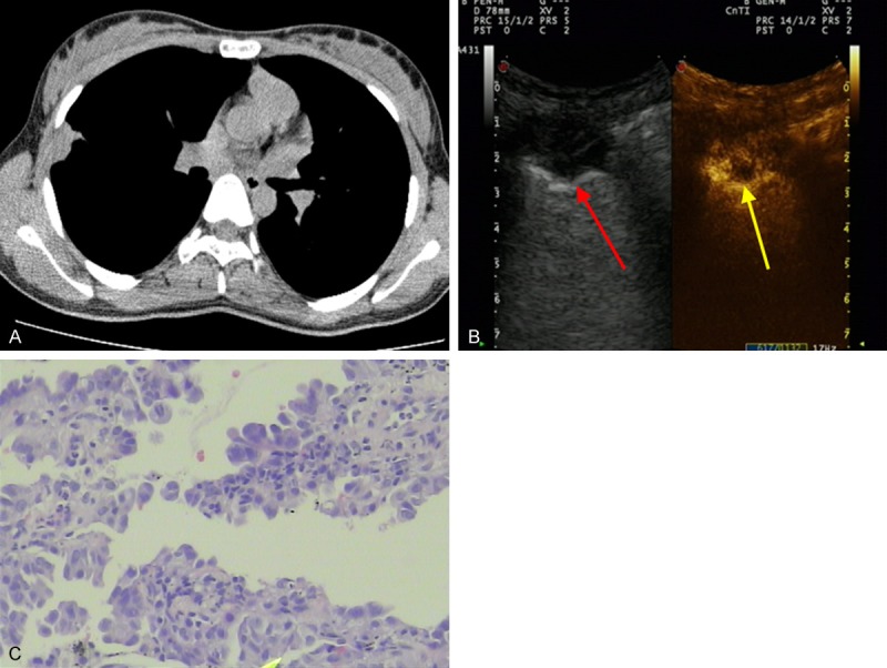
Figure 2.

Images of a 56-year-old man with chest pain for two weeks. A. Chest CT revealed a subpleural nodule in the middle lobe of the right lung (1.1 × 2.0 cm, arrowhead). B. Conventional US scan showed a subpleural nodule (red arrowhead) with a low echo texture; CEUS with SonoVue showed the enhanced area and necrotic area (yellow arrowhead). C. Biopsy sample obtained from the subpleural nodule showed adenocarcinoma cells (H&E staining; magnification, × 100).
