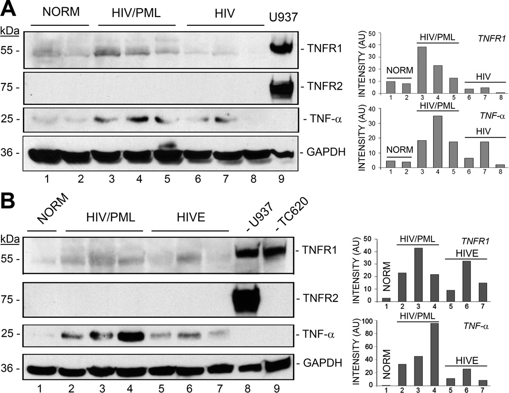
Figure 1. Analysis of expression of TNF-α and its receptors in HIV-1/PML clinical samples and controls by Western blot.
A. Brain tissues from HIV-negative control (NORM, n=2), HIV-1/PML (n=3), and HIV-positive patients with no CNS pathology (n=3) were analyzed for expression levels of TNF-α, TNFR1 and TNFR2 by Western blotting. The loading control was GAPDH. U937 human monocytic leukemia cell line lysate was used as a positive control for TNFR1 and TNFR2. Quantitation of the Western blot was performed and is shown in the histogram on the right with band intensities given in arbitrary units (AU). B. Brain tissues from a second set of clinical samples were analyzed: HIV-negative control (NORM, n=1), HIV-1/PML (n=3), HIV encephalitis (HIVE, n=3). U937 human monocytic leukemia cell line lysate was used as the positive control for TNFR1 and TNFR2and TC620 oligodendroglioma cell line lysate was used as a positive control for TNFR1.

