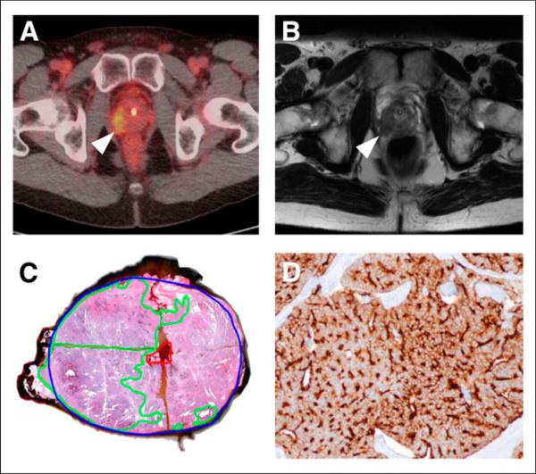
FIGURE 2.
Correlation between focal uptake in right lateral prostate apex (arrowhead) on 18F-DCFBC PET (A), abnormal low T2 signal (arrowhead) on MR imaging (B), and tumor on gross surgical pathology, as outlined in green (C). Pathologic specimen from same tumor shows strong immunohistochemical staining for PSMA (brown color) (D).

