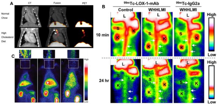
Figure 2.

Representative images using antibodies as radiotracers for atherosclerosis imaging. (A) In vivo PET/CT imaging in LDLR-/- mice injected with 64Cu-DOTA-anti-P-selectin mAb, demonstrating signal uptake in the brachiocephalic artery and aortic arch, as indicated by arrowhead and arrow, respectively. (B) Planar images after injection 99mTc-LOX-1-mAb compared to 99mTc-IgG2a in WHHL-MI and control rabbits. White arrows indicate aorta, K is kidney, L is liver, S is spleen. (C) Noninvasive planar images with injection of 99mTc-chP3R99 mAb by atherosclerotic (left image) and healthy (center image) rabbits, demonstrating the accumulation in the carotid indicated by arrowheads, compared to the injection of control radiotracer 99mTc-chT3 mAb (right image). Modified with permission [39, 41, 42].
