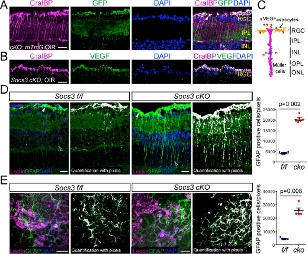
Figure 6. Neuronal/glial deficiency of Socs3 results in activated Müller glial cells and astrocytes.
(A) In P17 Socs3 cKO;mTmG reporter retinas with OIR, CralBP, labeled Müller cells and astrocytes colocalized with GFP. Cross-sections were stained for Müller cells and astrocytes with anti-CralBP (magenta) and cell nuclei with DAPI (blue). White arrow: astrocytes or end-feet of activated Müller cells. Open arrowheads: activated Müller cells. Scale bar: 10 µm. Images are representative of 3 mice per group. (B) Cross-sections from P17 Socs3 cKO retinas with OIR were stained with CralBP (magenta), VEGF (green) and DAPI (blue). VEGF colocalized with CralBP positive cells. Scale bar: 10 µm. (C) A diagram showing VEGF in the RGC and INL layers including activated astrocytes and Müller cells. (D) In P17 retinas with OIR, GFAP-labeled activated Müller cells in Socs3 cKO retinas were increased compared to Socs3 f/f control retinas. Cross-sections from P17 Socs3 cKO and Socs3 f/f control retinas with OIR were stained for endothelial cells with isolectin B4 (magenta), activated Müller cells and astrocytes with anti-GFAP (green) and cell nuclei with DAPI (blue). Scale bar: 25 µm. (E) GFAP-labeled astrocytes and end-feet of activated Müller cells in flat-mounted P17 Socs3 cKO and Socs3 f/f retinas with OIR. Representative images in A, B, D, and E were from 6 mice per group. White arrow: astrocytes or end-feet of activated Müller cells. Open arrowheads: activated Müller cells. Scale bar: 50µm.

