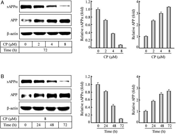
Figure 1.
Western blotting and quantification of the APP-α-processing after the ACAT1 inhibition (A) The human SK-N-SH cells were treated with the CP at different concentrations (0, 2, 4, or 8 µM) for 72 h. The immunoblotting of the cultured media and cell lysates as well as the quantification of the sAPPα or APP protein were performed according to the procedures described in the ‘Materials and Methods’. The data were the mean ± SD from three quantifications. The relative levels of proteins were expressed as fold to the controls without the CP treatment. (B) The human SK-N-SH cells were treated with 8 µM CP for different time intervals (0, 24, 48, or 72 h). The data were obtained by the same methods as indicated in (A).

