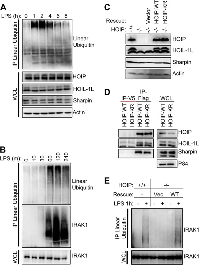
FIG 2 .

Kinetics of LUBAC activity and complementation of HOIP-deficient cells. (A) Mouse A20.2J B cells were stimulated with 10 µg/ml LPS to activate TLR4 signaling and induce LUBAC enzymatic activity. Cellular linear-ubiquitin levels were determined by immunoprecipitation with a linear-ubiquitin-specific antibody. Resting cells have low levels of linear ubiquitin relative to cells stimulated with LPS for 1 h. (B) As for panel A but with earlier time points as well as immunoblotting for IRAK1. (C) Mouse A20.2J HOIP−/− cells were complemented with vector, HOIP-WT, or HOIP-KR by lentiviral infection. LUBAC expression in complemented cells was compared to that in A20.2J HOIP+/+ cells by immunoblot analysis. (D) HOIP-Flag was immunoprecipitated from lysates of cell lines generated for panel A using Flag or IgG control antibody to determine LUBAC formation. (E) To verify function of complement cell lines, cells were stimulated with 10 µg/ml LPS for 1 h and then lysed in LUIP buffer. A linear-ubiquitin-specific antibody was used to precipitate linear ubiquitin chains, followed by immunoblotting with anti-IRAK1. Data are representative of three independent experiments.
