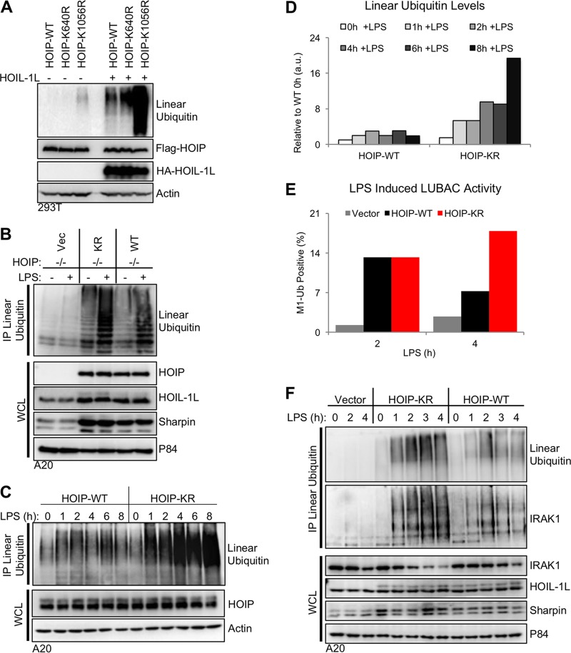
FIG 4 .
Mutation of HOIP lysine 1056 increases LUBAC enzymatic activity. (A) HOIP constructs were transfected into human 293T cells in the presence or absence of HOIL-1L. At 24 h after transfection, cells were lysed, and LUBAC protein expression and linear-ubiquitin-chain levels were determined by immunoblot analysis of cell lysates. (B) A20.2J HOIP−/− complemented cells from Fig. 2C were stimulated with 10 µg/ml LPS to activate TLR4 signaling and induce LUBAC enzymatic activity. Endogenous linear-ubiquitin levels were determined by immunoprecipitation with a linear-ubiquitin-specific antibody. (C) As described for panel B, but with LPS stimulation for various time points to compare linear-ubiquitin levels between WT and KR cells. (D) Densitometry analysis of the linear-ubiquitin immunoblot from panel C. All values are relative to WT cells prior to stimulation. (E) A20.2J HOIP−/− complemented cells stimulated as described for panel C were stained for linear ubiquitin using M1Ub-APC, and linear-ubiquitin-positive cells were quantified by flow cytometry. (F) A20.2J HOIP−/− complemented cells were stimulated as described for panel C. The immunoblot for IRAK1 shows the presence of linear ubiquitin chains on IRAK1 upon TLR4 activation. Data are representative of three independent experiments.

