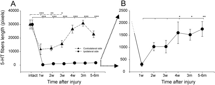
Fig 6. Serotonin fiber length.
(A) Serotonin fiber length (in pixels) at the lumbar spinal cord level (below the area of hemisection) in the ipsi- and contralateral sides before and at consecutive time points after injury (mean±SEM). (B) Expanded scale of the diagram in (A) to show changes in serotonin fiber length (in pixels) in the lumbar spinal cord level (below the area of hemisection) in the ipsilateral side at consecutive time points after the injury (mean±SEM). Abbreviations: w—weeks post operation, m—months post operation. * − p<0.05, ** − p<0.01, *** − p<0.001.

