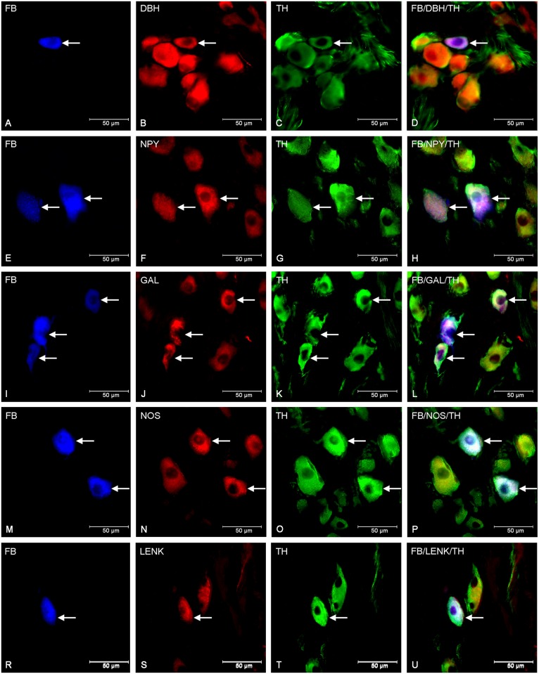
Fig 3. Immunohistochemical characteristic of traced neurons in animals of ASA group.
Fluorescent micrographs showing changes in immurectivity of FB-labelled neurons (A, E, I, M, R) in the porcine CCMG of ASA- treated animals. Photographs C, G, K, O and T show neurons immunoreactive to TH and simultaneously to DβH (B), NPY (F), GAL (J), nNOS (N) and LENK (S). Photographs D, H, L, P, and U have been created by digital superimposition of three colour channels. Single perikarya containing TH/DβH (B, C) in contrast to two in control group (Fig 1B and 1C) were visible. Moreover increase in the number of GAL- IR (J) and NPY-IR (F) neurons was observed and the novo-synthesis of nNOS (N) and LENK (S) was detected.

