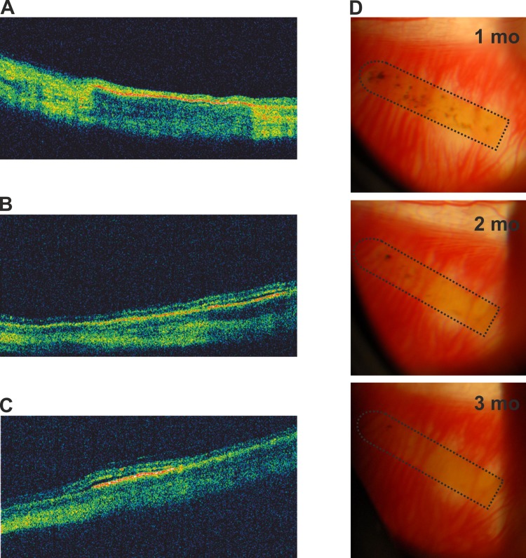
Fig 5. In vivo follow-up of hESC-RPE-PI implanted rabbit eyes.
OCT scans showing hESC-RPE-PI in three different rabbit eyes three months after transplantation (A-C). Representative fundus photographs showing hESC-RPE-PI (marked with dotted line) of one rabbit one, two and three months after transplantation (D).

