Fig. 2.
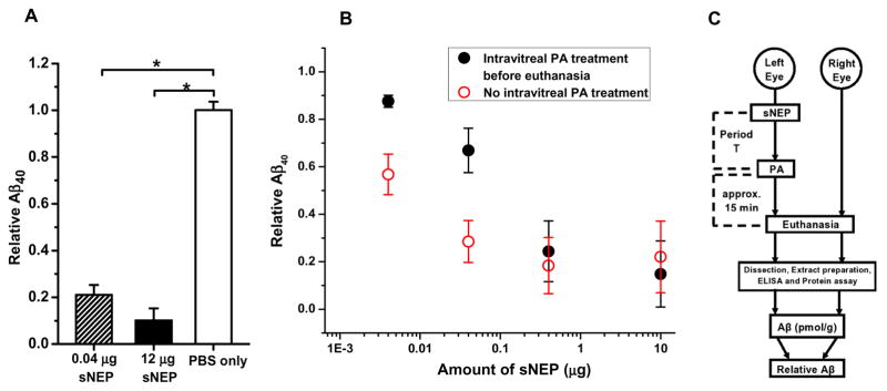
Aβ40 cleavage in C57BL/6J eyes by intra-vitreally delivered sNEP. A: Relative Aβ40 at 2 hr post-treatment in mice for which treatment consisted PBS alone (n=2; open bar), 0.04 μg sNEP (n=2; shaded bar), or 12 μg sNEP (n=2; filled bar). Asterisks indicate statistical significance with p<0.05. Intra-vitreal delivery of PBS alone (open bar) had no significant effect on relative Aβ40 (p>0.7). B: Results obtained with differing concentrations of delivered sNEP with PA treatment (2 μL of 1 mM PA) (filled circles) and without PA treatment (open circles). Each data point indicates results obtained from n ≥ 3 animals. C: Diagram illustrating the adopted, routine in vivo treatment of the test (left) eye and subsequent procedures. “Period T” refers to the post-sNEP-treatment period.

