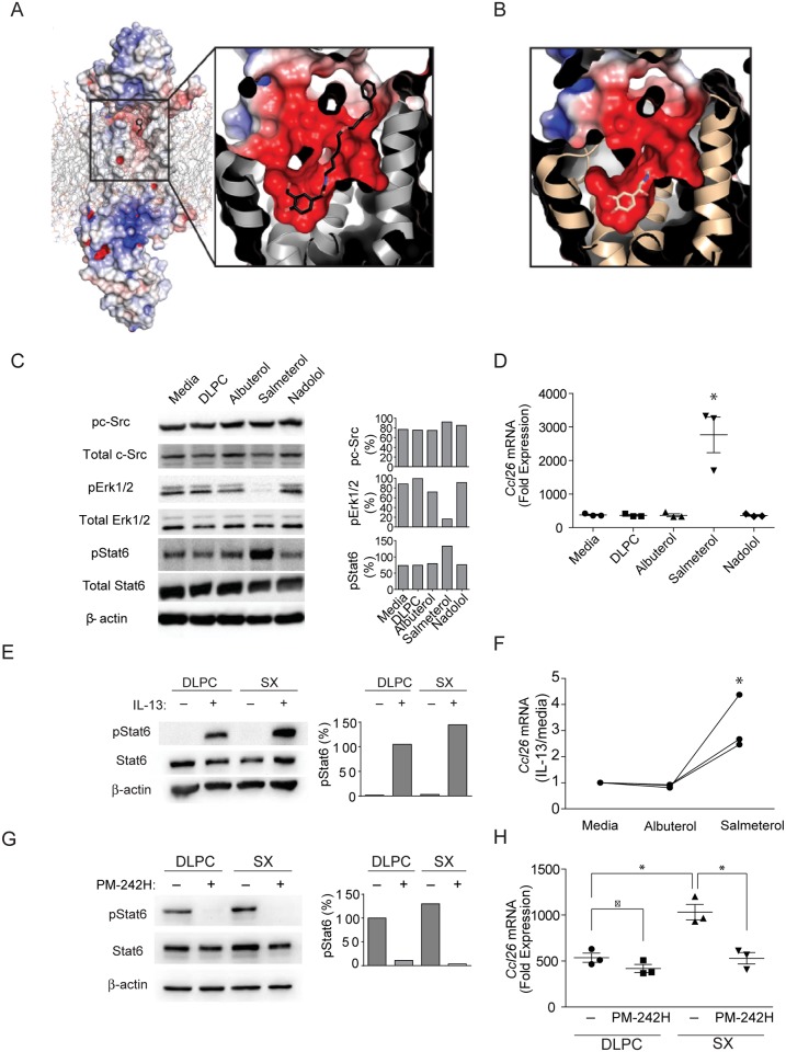
Fig 2. Salmeterol potentially binds aberrantly to the β2-AR active site and alters signaling function.
(a) Model of the β2-AR bound to salmeterol. Salmeterol is docked to the β2-AR bound to hydroxybenzyl isoproterenol (PDB Code: 4LDE) and energy minimized using Coot [62]. The entire model is shown with electrostatic surface potential calculated with APBS [38] on the left and a zoom in view of the binding pocket is shown on the right. Salmeterol is colored by atom type, with carbon colored black, nitrogen colored blue, and oxygen colored red. (b) Similar rendering of the β2-AR binding pocket with adrenaline bound [48], but with the carbon atoms of adrenaline colored yellow. (c-h) A549 human airway epithelial cells were exposed to IL-13 for 5 days and then to DLPC liposome vehicle, albuterol, salmeterol, or nadolol for 5 days after which (c) total and phosphorylated signaling proteins were determined in relation to beta actin as indicated. (d) Induction of Ccl26 mRNA as assessed by real time qPCR was determined under similar conditions in A549 cells. *: P < 0.05 determined by ANOVA (n = 3 technical replicates). (e) Total and phosphorylated STAT6 and (f) Ccl26 mRNA were quantitated from IL-13-stimulted human bronchial epithelial cells that were exposed to salmeterol or albuterol as indicated. *: P < 0.05 determined by ANOVA (n = 3 technical replicates). The effect of the phosphopeptidomimetic PM-242H on (g) STAT6 phosphorylation and (h) Ccl26 mRNA expression were further determined in IL-13-stimulated A549 cells. *: P < 0.05 determined by Mann Whitney test. Data are from one of 3 or more independent and comparable biological experiments.

