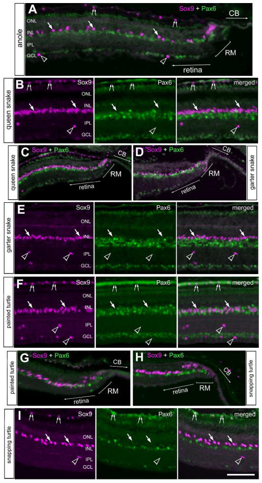
Figure 2.
Sox9 and Pax6 are expressed by cells in central and peripheral regions of the retina. Sections of peripheral (A,C,D,G,H) and central (B,E,F,I) retina were labeled with antibodies to Sox9 (magenta) and Pax6 (green). Retinas were obtained from the eyes of anoles (A), queen snake (B,C), garter snake (D,E), painted turtle (F,G) and snapping turtle (H,I). Arrows indicate the nuclei of Müller glia, small double-arrows indicate the nuclei of RPE cells, and hollow arrow-heads indicate presumptive NIRG-like cells. The scale bar (50 μm) in panel C applies to C–E, the bar J applies to H–J and the bar in O applies to A,B,F,G,K and O. Abbreviations: ONL – outer nuclear layer, INL – inner nuclear layer, IPL – inner plexiform layer, GCL – ganglion cell layer, RM –retinal margin, CB – ciliary body.

