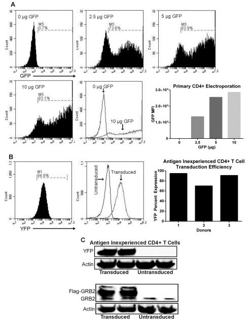
Figure 1. Electroporation of activated T cells and transduction of antigen inexperienced primary human CD4+ T cells.
(A) Activated primary human CD4+ T cells were electroporated with various concentrations of pmaxGFP plasmid using Lonza Amaxa electroporator, and cells expressing GFP were detected after 24 hrs. Bottom right: Quantification of GFP MFI on electroporated cells. Data represents 3 independent experiments with similar results (B) Left: antigen inexperienced primary CD4+ were transduced with lentiviruses expressing LUC shRNA for 4 days and then YFP expression was measured. Right: Percent YFP expression of transduced antigen inexperienced CD4+ T cells compiled from three donors. (C) Antigen inexperienced primary CD4+ T cells were transduced for 4 days with lentiviruses carrying YFP or Flag-GRB2 constructs. Cells were lysed and protein expression of YFP, GRB2, and actin were detected via immunoblotting. Data represents 3 independent replicates with similar results.

