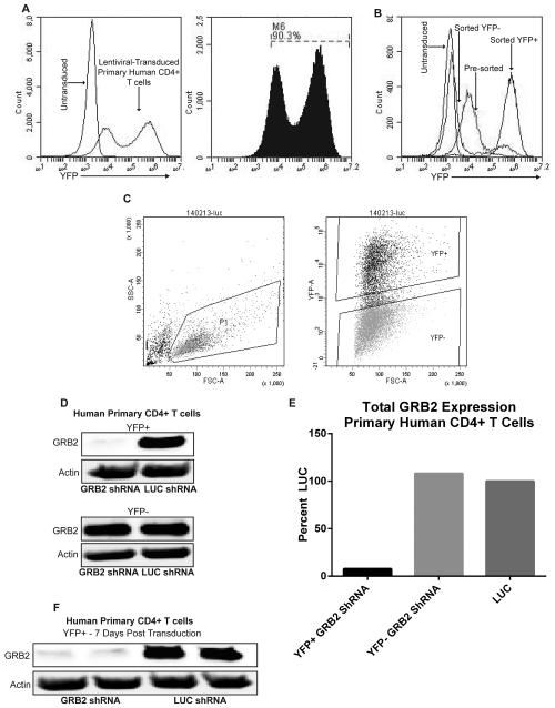
Figure 2. Transduction of activated primary human CD4+ T cells using lentiviruses.
(A) Flow cytometry analysis of activated primary human CD4+ T cells that were transduced for 3 days with pLK4A lentiviral vectors carrying Luciferase (LUC) shRNA and YFP. Data represents 4 independent experiments with similar results. (B) Primary human CD4+ T cells were transduced with pLK4A lentiviral vectors expressing YFP along with LUC shRNA; illustrated groups are: untransduced, 3 day transduction pre-sort, Post-Sort YFP+, Post-Sort YFP−/low. (C) Left: Gate on live primary human CD4+ T cells transduced with pLK4A expressing YFP and LUC shRNA. Right: Gates for sorting YFP+ and YFP− populations. Data represents 3 independent replicates with similar results. (D) Primary human CD4+ T cells transduced with shRNAs against GRB2 or LUC were sorted into YFP+ and YFP− fractions after 3 days of transduction, lysed and probed for GRB2 expression. (E) Quantification of immunoblots from “D”. (F) Immunoblot of GRB2 expression from “D” 7 days post transduction.

