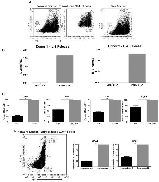
Figure 5. Transduced YFP+ are highly active and larger in size compared to YFP− primary CD4+ T cells.
(A) Activated primary CD4+ T cells were transduced with pLK4A lentiviruses carrying LUC shRNA and YFP for 3 days and then probed for YFP expression and forward/side scatter; S/L indicates Small/Large fractions. (B) Primary human CD4+ cells were transduced with YFP+ pLK4A lentiviruses expressing LUC shRNA. Sorted YFP+/- cells were stimulated with 5 µg/mL anti-CD3 coated plates for 24 hrs. The supernatants were probed for IL-2 production via ELISA. Illustrated is IL-2 production data from CD4+ primary cells obtained from two different donors. (C) Cells from “A” were surface stained with CD44 or CD69 conjugated with Alexa-fluor 647 and then gated on YFP−, S/L YFP+, and L YFP+ and then MFI was collected from 100,000 live gate events. Groups were graphed as ±SEM of YFP+ cells as indicated from three independent donors. (D) Untransduced activated primary CD4+ cells from the same donors as in “C” were stained with surface CD44 or CD69 conjugated with Alexa-fluor 647 and then gated on large “L” and small “S” fractions. Data was graphed as ±SEM of large cells from three independent donors.

