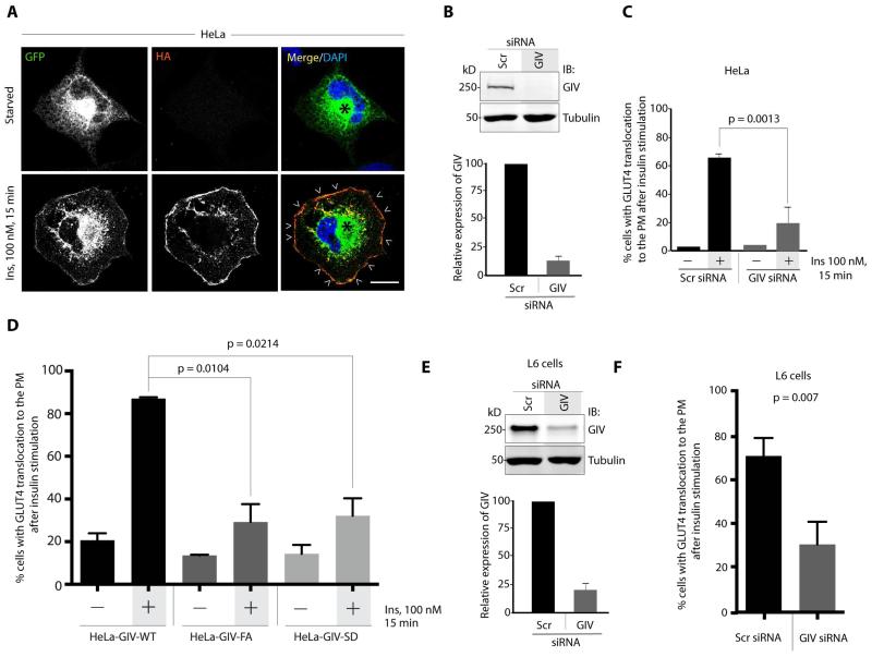
Figure 1. GIV’s GEF function is essential for GLUT4 exocytosis.
(A) Starved or insulin-stimulated HeLa cells expressing HA-GLUT4-GFP were fixed, stained with HA mAb and analyzed by confocal microscopy. Exocytosed GLUT4 at the PM (arrowheads) was detected by surface labeling with HA (red), whereas total GLUT4 was detected by GFP signal. Bar = 10 μm. (B) Top: Lysates of HeLa cells treated either with control (Scr) or with GIV siRNA were analyzed for GIV and tubulin by immunoblotting (IB). Bottom: Bar graph displays efficiency of GIV depletion. (C) Control (Scr) and GIV-depleted (GIV siRNA) HeLa cells were analyzed for insulin-triggered exocytosis of HA-GLUT4-GFP by confocal microcoscopy as in A. Bar graph displays % cells with exocytosed GLUT4. Error bars represent mean ± S.D. n = 3. (D) HeLa cells stably expressing siRNA-resistant GIV-WT, GIV-FA or GIV-SD were depleted of endogenous GIV by siRNA, serum starved and subsequently analyzed for exocytosis of HA-GLUT4-GFP after insulin stimulation as in A. Bar graph displays % cells with exocytosed GLUT4. Error bars represent mean ± S.D. n = 3. (E) Top: Lysates of L6 cells treated either with control (Scr) or with GIV siRNA were analyzed for GIV and tubulin by immunoblotting (IB). Bottom: Bar graph displays efficiency of GIV depletion. (F) Control (Scr) and GIV-depleted (GIV siRNA) L6 cells were analyzed for insulin-triggered exocytosis of HA-GLUT4-GFP by confocal microcoscopy as in A. Bar graph displays % cells with exocytosed GLUT4. Error bars represent mean ± S.D. n = 3.

