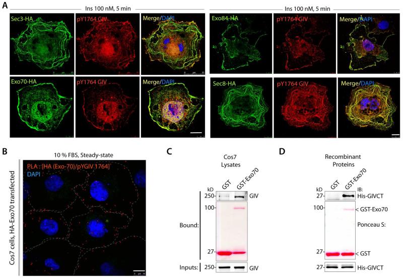
Figure 3. GIV colocalizes with the exocyst complex and directly binds the subunit Exo-70.
(A) Tyrosine phosphorylated GIV colocalizes with several subunits of the exocyst complex. Serum starved Cos7 cells expressing various subunits of the exocyst complex, as indicated were stimulated with insulin, fixed and subsequently stained for pY1764-GIV (red), HA (green) and DAPI/DNA (blue). Bar = 10 μm. (B) Cos7 cells expressing Exo70-HA were grown in 10% FBS (steady-state; right) and analyzed for interaction between GIV and Exo-70 by in situ PLA using rabbit anti-pY1764-GIV and mouse anti-HA. Red dots = interaction. Bar = 10 μm. (C-D) Pulldown assays were carried out using either Cos7 cell lysates (C) or recombinant His-GIV-CT (aa 1660-1870) (D) as source of GIV and GST or GST-Exo70 immobilized on glutathione beads. Inputs and bound proteins were analyzed by immunoblotting (IB) for GIV (left) or for His (His-GIV-CT, right). Equal loading of GST proteins was confirmed by Ponceau S stain.

