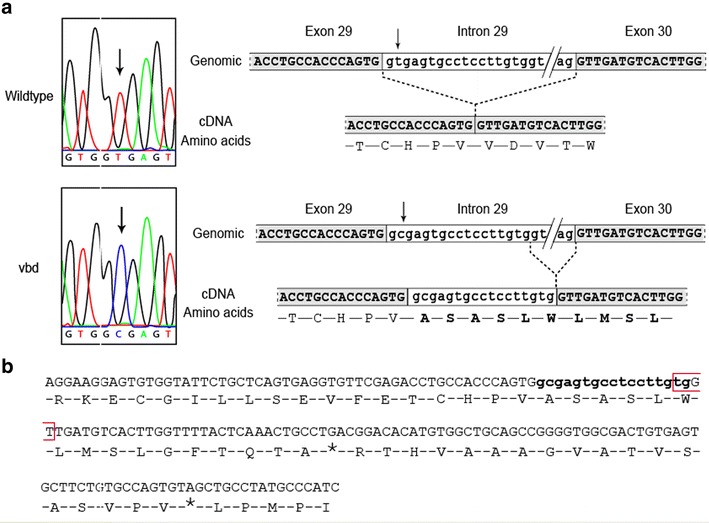
Fig. 4.

Sequence analysis spanning the vbd mutation site. a (Left) Sequence chromatograph of the otog gene at the level of the splicing site exon 29/intron 29 showing the T > C transition (arrows); (right) partial genomic sequence of the region spanning exon 29 to exon 30 with the corresponding cDNA and amino acid sequence. Top: wildtype; bottom: Otog vbd/vbd mutant. b cDNA sequence surrounding the vbd mutation showing the insertion of 17 bp (lowercase bases in bold) including a new splicing site (open red box). The resulting reading frame shift leads to a premature stop codon in the amino acid sequence between VWFD 3 and VWFD 4 domain (uniprot)
