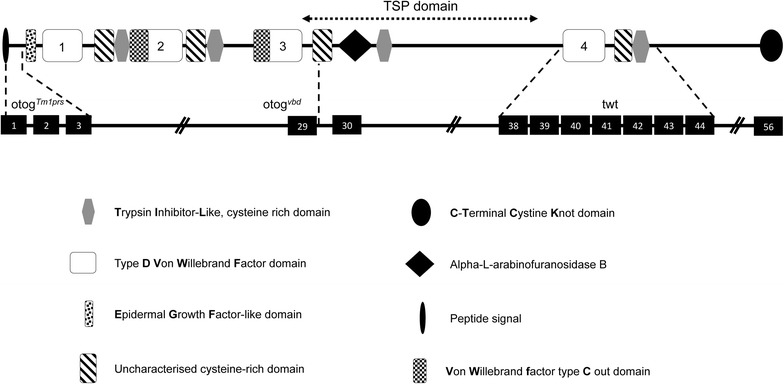
Fig. 5.

Schematic representation of OTOG protein. The emplacement of the different domains of OTOG protein are shown (top). The positions of the three mutations and the corresponding exons where the mutations occur are shown at the bottom. otogvbd (T > C transition at the level of the splicing site exon 29/intron 29 of the otog gene), otogtm1prs (replacement of the majority of exon 1 and all of exons 2 and 3 by the lacZ gene fused in frame with the translation initiation codon of Otog), and twister (spontaneous recessive mutation showing a discrete rearrangement within the 3′ part of the Otog locus, between exons 38 and 44 not precisely identified)
