Figure 12.
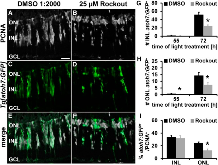
Rock inhibition affects neuronal lineage commitment of ONL, but not INL, proliferative cells. A–F, Single z-plane confocal images of light-damaged albino;Tg[atoh7:GFP]rw021 zebrafish that were systemically exposed to either DMSO (A, C, E) or 25 μm Rockout (B, D, F) and immunocytochemically labeled for PCNA (A, B, E, F) and EGFP (C–F) show fewer atoh7:GFP-positive cells in Rockout-treated retinas relative to DMSO controls at 72 h of light treatment. Histogram depicting the number of atoh7:GFP-positive cells in the INL (G) and ONL (H) at 55 and 72 h of light treatment. I, Histogram displaying that the percentage of PCNA-positive cells that express atoh7:GFP in the ONL was significantly reduced, but not in the INL, of Rockout-treated retinas compared with DMSO controls at 72 h of light treatment. GCL, Ganglion cell layer. Data are shown as mean ± SE, n ≥ 18, Student's t test,*p < 0.05. Scale bar, 20 μm for A–F.

