Figure 14.
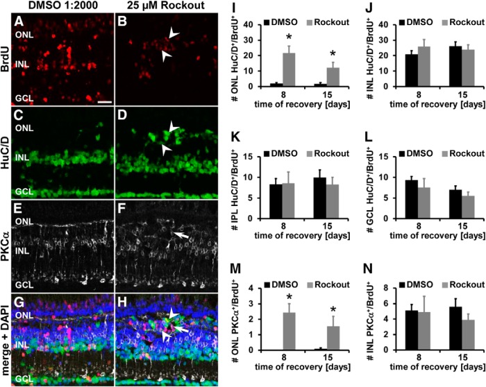
Increased generation of HuC/D- and PKCα-positive neurons in the basal ONL in Rock-inhibited retinas. A–H, Single z-plane confocal images of light-damaged albino zebrafish that were systemically exposed to either DMSO (A, C, E, G) or 25 μm Rockout (B, D, F, H) from 28–72 h of light treatment. Zebrafish intraperitoneally injected with BrdU were allowed to recover for 8 d and were immunocytochemically labeled for BrdU (A, B, G, H), HuC/D (C, D, G, H), and PKCα (E–H). Rock inhibition caused an accumulation of HuC/D- and PKCα-positive cells in the basal ONL region and a subset of these cells colabeled with BrdU (arrowheads and arrow, respectively). I–N, Histograms depicting the number of HuC/D and BrdU double-positive cells in the ONL (I), INL (J), IPL (K), and ganglion cell layer (GCL; L) and the number of PKCα and BrdU double-positive cells in the ONL (M) and INL (N) at 8 and 15 d of recovery in retinas exposed to DMSO or Rockout from 28–72 h of light treatment. Data are shown as mean ± SE, n ≥ 12, Student's t test,*p < 0.05. Scale bar, 20 μm for A–H.

