Figure 6.
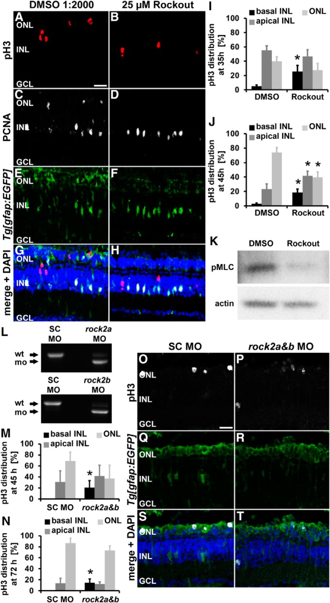
Inhibition of Rocks as well as rock2a and rock2b double knockdowns alter the position of pH3-positive Müller glia and of NPC nuclei after light damage. A–H, Single z-plane confocal images of albino;Tg[gfap:EGFP]nt11 zebrafish that were exposed to either DMSO (A, C, E, G) or 25 μm Rockout (B, D, F, H) were immunocytochemically labeled with pH3 (A, B) and PCNA (C, D). Inhibition of Rocks altered the position of pH3-positive Müller glia (A, B, G, H), but not the number of PCNA-positive cells (C, D, G, H) at 35 h after starting the light treatment. Histograms depict the distribution of pH3-positive nuclei in the ONL and apical and basal INL in DMSO- and Rockout-treated zebrafish at 35 h (I) and 45 h (J) after starting the light treatment. Data are shown as mean ± SE, n ≥ 18, Student's t test, *p < 0.05 between corresponding retinal regions in DMSO- and Rockout-treated samples. K, Immunoblot of phosphorylated MLC (pMLC, Thr18/Ser19) and actin as a loading control in DMSO and Rockout-treated retinal lysates. n = 3. L, RT-PCR was performed on cDNA obtained from embryos to determine the efficacy of the rock2a [top, 24 h postfertilization (hpf)] and rock2b (bottom, 72 hpf) splice junction morpholinos, respectively. M, N, Histograms of the distribution of pH3-positive nuclei in the retina depicting that significantly more pH3-positive nuclei were located in the basal INL in rock2a and rock2b double morphants relative to standard control morphants at 45 h (M) and 72 h (N) of light treatment. O–T, Single z-plane confocal images of albino;Tg[gfap:EGFP]nt11 zebrafish that were electroporated with either a standard control morpholino (O, Q, S; SC MO) or a combination of rock2a and rock2b splice site morpholinos (P, R, T), light-damaged for 72 h, and immunocytochemically labeled for pH3 (O, P, S, T) and GFP (Q–T) and counterstained with the nuclear dye DAPI (S, T). GCL, Ganglion cell layer; SC MO, standard control morpholino; wt, wild-type. Scale bar, 20 μm for A–H and O–T.

