Figure 7.
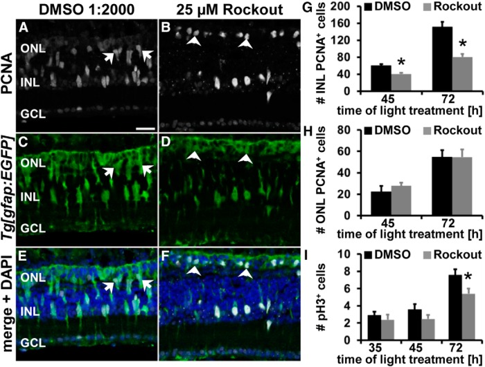
Rock inhibition reduces NPC proliferation without affecting the number of mitotic cells. A–F, Single z-plane confocal images of light-damaged albino;Tg[gfap:EGFP]nt11 zebrafish that were exposed to either DMSO (A, C, E) or 25 μm Rockout (B, D, F) and immunocytochemically labeled with PCNA (A, B) and EGFP (C–F) show reduced numbers of PCNA-positive cells at 45 h of light treatment. Arrows and arrowheads indicate PCNA-positive ONL nuclei with an elongated (A, C, E) or a round morphology (B, D, F), respectively. Histogram depicting the number of PCNA-positive cells in the INL (G) and ONL (H) in DMSO- and Rockout-treated retinas at 45 and 72 h of light treatment. I, Histogram displaying the number of pH3-positive cells in Rockout- and DMSO-exposed retinas at 35, 45, and 72 h after starting the light treatment. GCL, Ganglion cell layer. Data are shown as mean ± SE, n ≥ 12, Student's t test, *p < 0.05. Scale bar, 20 μm for A–F.

