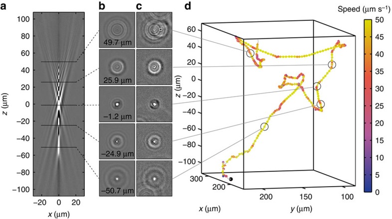
Figure 2. Tracking bacteria in 3D by comparing their out-of-focus diffraction patterns to a reference library.
(a) A vertical slice through a reference library created by combining 73 aligned image stacks obtained for 1 μm silica beads. (b) Horizontal slices from the reference library at positions marked in a. (c) Images of a swimming E. coli bacterium at the corresponding positions. (d) Reconstructed 3D trajectory for the bacterium in c (see Supplementary Movie 3 for a rotating view). The trajectory starting point is marked by a black dot.

