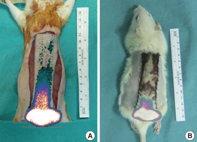
Fig. 6. Combination of a scintigram and a photograph.

In the control group, (A) half of the flap was perfused before the experiment, and (B) perfusion was only found in the pedicle region after the experiment.

In the control group, (A) half of the flap was perfused before the experiment, and (B) perfusion was only found in the pedicle region after the experiment.