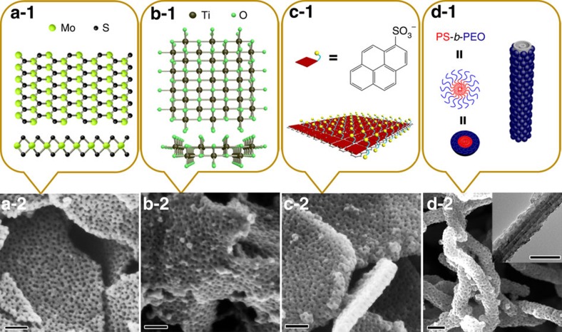
Figure 4. Patterning of various functional free-standing surfaces.
(a-1) Structure of MoS2 nanosheets in top view (upper) and side view (lower). (a-2) SEM image of large-pore mesoporous PPy nanosheets on MoS2 nanosheets. (b-1) Structure of titania nanosheets in the [010] (upper) and [001] (lower) directions. (b-2) SEM image of large-pore mesoporous PPy nanosheets on titania nanosheets. (c-1) Illustration of the 1-PSA-modified EG surfaces. (c-2) SEM image of large-pore mesoporous PPy nanosheets on EG nanosheets following modification with 1-PSA. (d-1) Illustration of CNTs wrapped by PS-b-PEO micelles. (d-2) SEM image of large-pore mesoporous PPy nanosheets on CNTs (inset of d is TEM image; scale bar: 100 nm).

