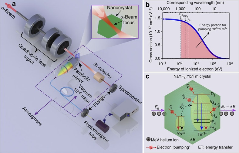
Figure 1. Experimental setup and proposed ionoluminescence mechanism.
(a) Artist's view of the basic experimental setup. The focused beam with a spot size of sub-30 nm features can be achieved using a spaced triplet of compact magnetic quadrupole lenses. A Si surface barrier detector is equipped for measuring the energy loss distribution of the ions. (b) Calculated energy distribution of the ionized electrons by bombarding the MeV α-particles on the lanthanide-doped nanocrystals, showing different cross-sections of the resulting electrons at specific energies. Note that most of the ionized electrons have energies mainly located in the visible and infrared spectral region. (c) Proposed upconversion mechanism under α-beam irradiation. The incident helium ions with energy of E0 deposit a certain amount of energy (ΔE) onto the crystal to cause the atomic ionization inside the crystal. Subsequently, the ionized secondary electrons can release their energy, most likely during the electron-hole recombination process and successively transfer the energy to Yb3+ and Tm3+. An energy transfer from the excited Yb3+ to its neighbouring Tm3+ ions then populates the excited states (for example, 3H4, 1G4 and 1D2) of Tm3+.

