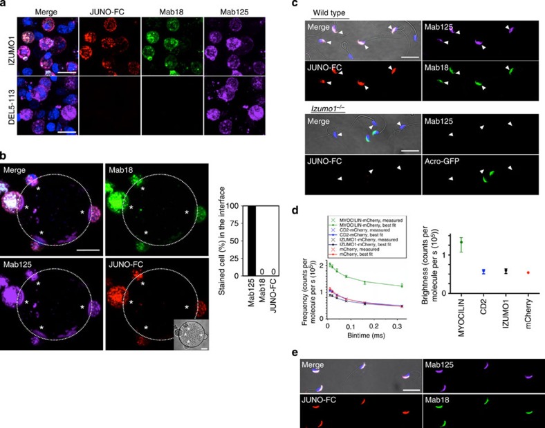
Figure 3. Recombinant JUNO binds to spermatozoon where IZUMO1 is monomeric.
(a) Binding of recombinant JUNO to IZUMO1. The JUNO-FC fusion protein labelled with α-mouse IgG antibodies-Alexa546 selectively bound to IZUMO1-expressing cells (red). Simultaneously, they were incubated with Mab18-Alexa488 (green) and Mab125-Alexa647 (magenta). (b) Cell–oocyte assay with recombinant JUNO. The same experiment was carried out with oocytes. The interface was devoid of JUNO-FC signal (asterisks). The right graph shows the percentage of stained cells in the cell–oocyte interface. Ten oocytes and fifty-seven attached cells were investigated in this analysis. The image of the oocyte is marked by dotted lines. Inset shows middle DIC image among Z-stacks. (c) Immunostaining of wild-type and IZUMO1-null spermatozoa with JUNO-FC. Wild-type fresh spermatozoa were incubated for 2 h in TYH medium with JUNO-FC (red), Mab18-Alexa488 (green) and Mab125-Alexa647 (magenta). Acrosome-reacted spermatozoa were detected with IZUMO1 antibodies (Mab18 and Mab125; shown by arrowheads). To detect the acrosome reaction in IZUMO1-null spermatozoa, Acro-GFP spermatozoa, which have green fluorescence that should disappear from the acrosome, were used to distinguish the acrosome reaction after 2 h of incubation in TYH medium with JUNO-FC and Mab125-Alexa647. Acrosome-reacted spermatozoa are shown by arrowheads. (d) IZUMO1-mCherry in spermatozoa showed monomeric brightness. The sperm lysate of IZUMO1-mCherry or COS-7 cell lysates expressing Myocilin-mChery, Cd2-mCherry or mCherry was prepared, and specific brightness of each protein was determined as described in the Methods. The true specific brightness was obtained from the bin time dependence (PCMH, left panel). The ranges of the upper and lower confidence interval (0.95) were also calculated from the five measurements and expressed as error bars of the mean. Brightness of IZUMO1-mCherry was statistically not different from that of mCherry or CD2-mCherry. Error bar represents CI95. (e) Immunostaining of acrosome-intact spermatozoa with JUNO-FC. Fresh spermatozoa collected from the epididymis of wild-type mice were mounted on glass slides, dried up and then immunostained with JUNO-FC (red), Mab18-Alexa488 (green) and Mab125-Alexa647 (magenta). Final concentration of all antibodies used was at 0.5 μg ml−1. JUNO-FC was added at 1 μg ml−1. Nuclei were stained with Hoechst 33342. Scale bar, 20 μm.

