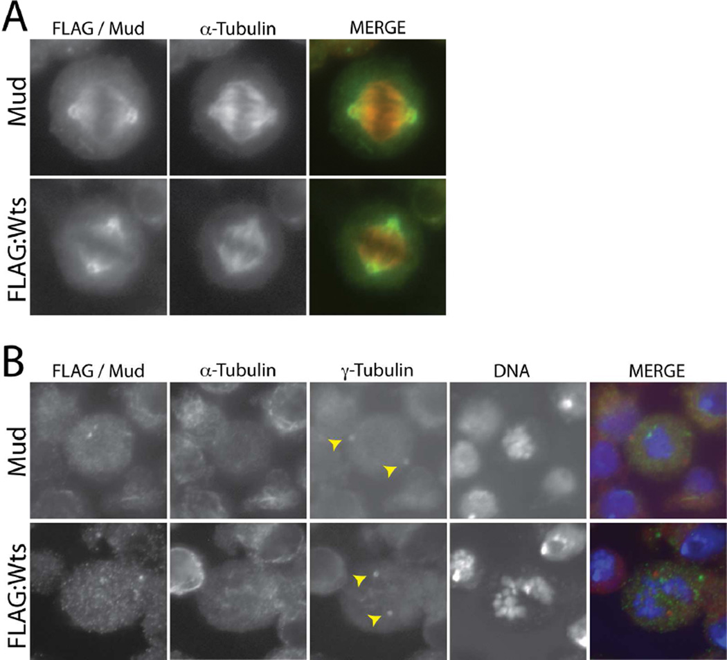
Figure 1. Warts localizes to mitotic spindle poles in mitotic S2 cells.
(A) Cells were transfected with full-length Warts tagged with an N-terminal FLAG epitope sequence (FLAG:Wts) and stained with antibodies against FLAG and α-tubulin. To visualize endogenous Mud, untransfected cells were stained with an α-tubulin and Mud antibodies. (B) To depolymerize spindle microtubules, cells were treated with colchicine (12.5 µM) for 2 hours prior to fixation and antibody staining. Yellow arrowheads indicate both γ-tubulin-positive centrosomes, to which neither Wts nor Mud show significant localization. α-tubulin staining indicates successful depolymerization of spindle microtubules; note this channel is not shown in the merge panel.

