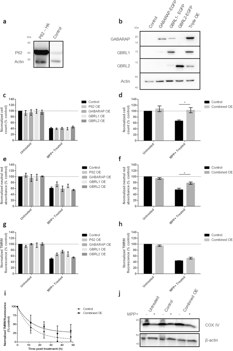
Figure 3. Overexpression of key autophagy proteins protects cells against MPP+ induced cell death.
(a,b) Overexpression of P62, GABARAP, GBRL1 and GBRL2 in BE(2)-M17 cells. Cells transfected with appropriate plasmids were harvested 72 h after transfection. Protein levels were measured by western blot and band density was normalised relative to actin. (c–h) Simultaneous overexpression of P62, GABARAP, GBRL1 and GBRL2 rescues MPP+ toxicity. Cells were transfected with expression constructs, treated with MPP+ (100 μM) and assayed. 4OE indicates simultaneous overexpression of P62, GABARAP GBRL1 and GBRL2. (c,d) Cell death was assessed by counting the number of morphologically normal cells in bright field images 48 h after MPP+ treatment. (e,f) Cell viability was assessed by neutral red uptake 36 h after MPP+ treatment. (g–i) Mitochondrial membrane polarisation was measured using TMRM fluorescence (g,h) 24 h after MPP+ treatment or (i) after 12, 24, 36 or 48 h MPP+ treatment. (j) Representative western blot of the mitochondrial protein COX IV 48 h after MPP+ treatment in the presence/absence of combined protein overexpression (n = 3). Bars represent mean values normalised to untreated control cells ± SEM (n = 3), data were analysed using a two way ANOVA with Sidak multiple comparison test * = P ≤ 0.05.

