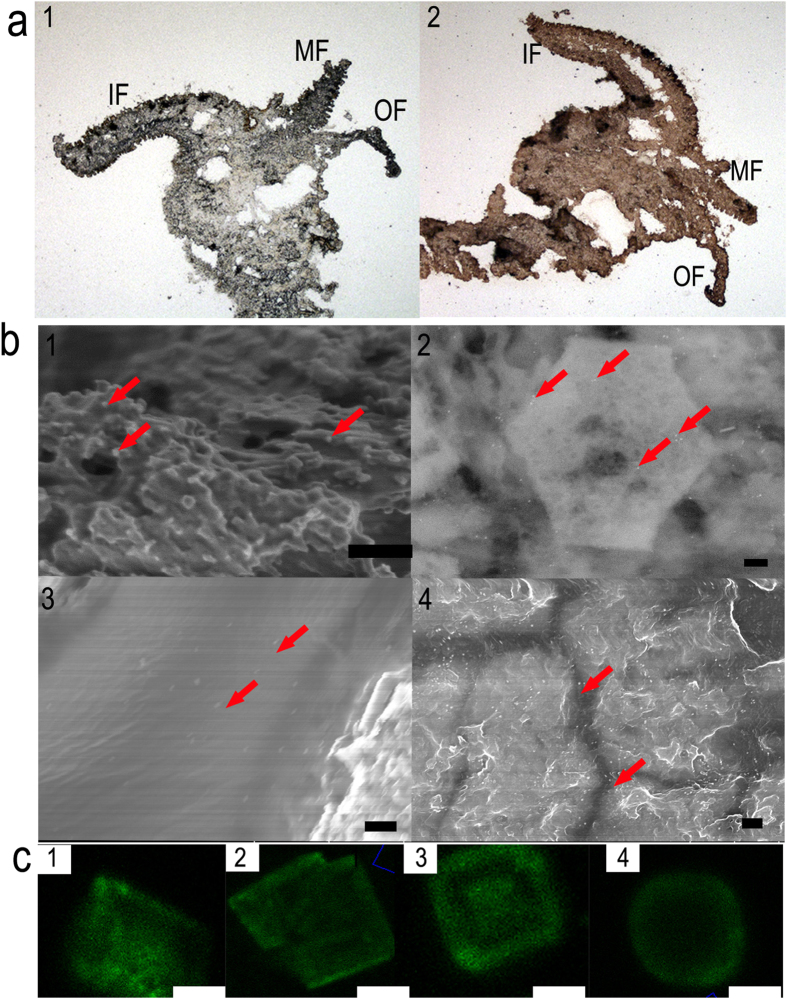
Figure 2. Immunolocalization of the shell matrix proteins (SMPs) of P. fucata.
A polyclonal antibody raised against mixed extracted proteins is used to identify EDTA-soluble matrices (ESMs) and EDTA-insoluble matrices (EISMs). (a) Immunohistochemical localization in the mantle epithelia: control without the first antibody (a1); mantle sections with the first antibody (a2) (MF: middle fold; OF: outer fold; IF: inner fold); (b) Immunogold labeling of SMPs on the EDTA-mounted prismatic and nacreous layers. Layers with the first antibody (b1-b4). b1, b3 are prismatic layers and b2, b4 are nacreous layers. The red arrowheads indicate gold nanoparticles. (Scale bars, 200 nm) (c) Confocal fluorescence laser scanning microscopy images of SMPs in vitro show synthetic calcite after immunolabeling. Calcite in the presence of 1 μg·mL−1 ESM-P (c1), EISM-P (c2), ESM-N (c3), EISM-N (c4). (Microscopy settings are identical. The control is shown in Figure S6. Scale bars, 10 μm.)

