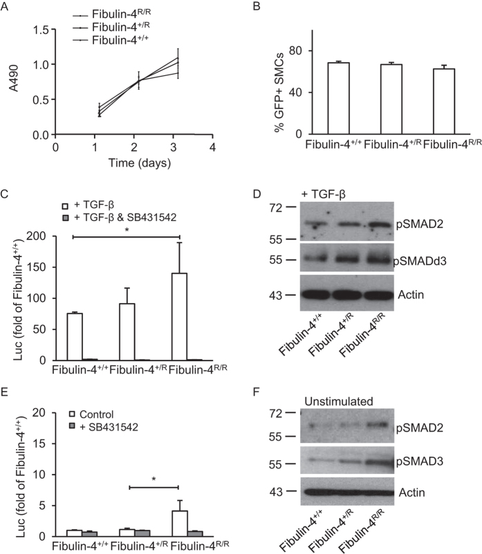
Figure 3. Increased TGF-β signalling in Fibulin-4 deficient SMCs.
(a) Fibulin-4+/+, Fibulin-4+/R and Fibulin-4R/R SMCs show similar proliferation rates in the time course of the experiment (MTS proliferation assay) (b) Transfection with green fluorescent protein (GFP) encoding plasmids display no difference in percentage of transfected SMCs between Fibulin-4+/+, Fibulin-4+/R and Fibulin-4R/R SMCs. (c) The TGF-β response assay reveals a gradual increase in TGF-β activity in Fibulin-4+/R and Fibulin-4R/R SMCs compared to Fibulin-4+/+ SMCs, after stimulation with exogenous TGF-β. Data represent fold change relative to unstimulated Fibulin-4+/+ SMCs. Addition of the ALK-5 kinase inhibitor (SB431542) abolishes the TGF-β response in Fibulin-4+/+, Fibulin-4+/R and Fibulin-4R/R SMCs. (d) Western blot analyses for TGF-β signalling downstream mediators pSMAD2 and pSMAD3 on TGF-β stimulated SMCs show a gradual increase in TGF-β signalling in Fibulin-4 deficient SMCs compared to Fibulin-4+/+ SMCs. (e) Measurement of basal TGF-β activity (no stimulation with exogenous TGF-β) by the CAGA-luciferase reporter show increased TGF-β activity in Fibulin-4R/R SMCs compared to Fibulin-4+/+ SMCs, which can be inhibited by the TβRI kinase inhibitor SB431542. (f) These data were confirmed by western blot analyses for pSMAD2 and pSMAD3. All data shown are representative for in total n = 3 independent experiments and all performed under serum starved conditions (*p < 0.05, **p < 0.01).

