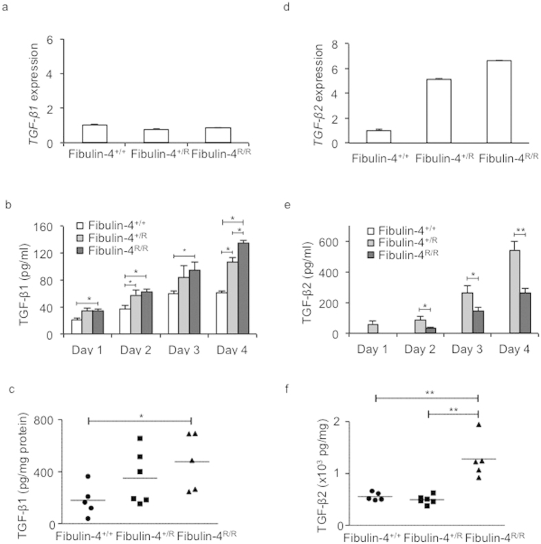
Figure 4. Strong increase of TGF-β2 levels in Fibulin-4 deficient SMCs.
(a) Fibulin-4+/R and Fibulin-4R/R SMCs show equal Tgf-β1 mRNA expression levels compared to Fibulin-4+/+ SMCs. (b) Increased TGF-β1 levels measured in conditioned medium (CM) from Fibulin-4R/R SMCs compared to Fibulin-4+/+ CM on day 1–4 after serum starvation. Fibulin-4+/R SMCs showed significant increased TGF-β1 levels on day 2 and 4 after serum starvation compared to Fibulin-4+/+ SMCs. Furthermore, on day 4 Fibulin-4R/R SMCs show significant increased TGF-β1 levels compared to Fibulin-4+/RSMCs (n = 4 per day for each genotype). Two-way ANOVA analysis for genotype and between days p < 0.05. (c) Gradually increased TGF-β1 is also observed in aortic arch lysates of Fibulin-4+/R (n = 6) and Fibulin-4R/R mice (n = 5) compared to Fibulin-4+/+ aortas (n = 5). This increase is significant in Fibulin-4R/R aortic arch lysates compared to Fibulin-4+/+ aortic arch lysates. (d) Fibulin-4+/R and Fibulin-4R/R SMCs show gradual increased Tgf-β2 mRNA expression levels compared to Fibulin-4+/+ SMCs. (e) Measurement of TGF-β2 revealed markedly increased levels in CM of Fibulin-4+/R and Fibulin-4R/R SMCs, while TGF-β2 was undetectable in CM of Fibulin-4+/+ SMCs (experiments were performed in at least 3 independent experiments). Two-way ANOVA analysis for genotype and between days p < 0.05. (f) Measurements in aortic arch lysates display significantly increased TGF-β2 in Fibulin-4R/R aortas compared to Fibulin-4+/R and Fibulin-4+/+ aortas (*p < 0.05, **p < 0.01).

