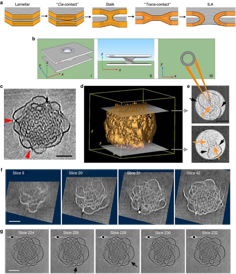
Figure 4. Membrane fusion initiates layer formation of bicontinuous cubic phase.
(a) Schematic of the membrane fusion process. (b) Model of the ILA (I), side (II) and top views (III). (c) Z-slice extracted from the ‘central' part of the tomogram showing membrane fusion events (red arrows). (d) 3D rendering of the whole volume of the cubosome with the position of the two top and bottom extreme slices of the shell (e), where ILAs (orange arrows) and the curved lamellar layers (black arrows) are visible. (f) The sequences extracted from the tomogram shows at the beginning only few ILAs close to the interface (slice 9). In the successive slices, the number of ILAs increases presenting an irregular organization (slice 20). At the centre of the particle, the packing of the ILA in the deep layers forms the bicontinuous cubic structure in which the water channels are well organized (slice 42). Slices were cut normal to the ILA axis. (g) Sequences of slices extracted from a tomogram of a small cubosome where visible ILAs are oriented at 90° (black arrows) from the ones of Fig. 4e,f. All scale bars, 50 nm.

