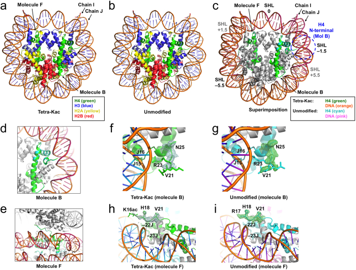
Figure 2. Crystal structures of the H4-tetra-acetylated NCP.
(a) The H4-K5/K8/K12/K16-tetra-acetylated NCP. (b) The unmodified NCP. (c) Superimposition of the two NCPs. Color codes for H4 and DNA are indicated at the bottom. The possible position for the molecule-B H4 tail around SHL –1.5 is shown by a dashed blue circle. The positions of the SHLs, at –5.5, –1.5, 0, +1.5, and +5.5, are indicated. SHLs +1.5 and +5.5, which are located behind the two DNA duplexes, are indicated in gray. (d) Superimposition of the molecule-B H4 structures. (e) Superimposition of the molecule-F H4 structures (horizontal view). The symmetrically-related NCP is depicted in gray. (f,g) Close-up views of the molecule-B N-terminal region in the H4-tetra-acetylated (f) and unmodified (g) NCPs. Both meshes indicate the 2mFo-DFc electron density maps corresponding to residues V21–I26. Residues 1–20 are disordered. (h,i) Horizontal views of the molecule-F N-terminal region in the H4-tetra-acetylated (h) and unmodified (i) NCPs. The meshes correspond to residues K16ac–V21 and R17–V21, respectively. All maps are contoured at 1.5 σ.

