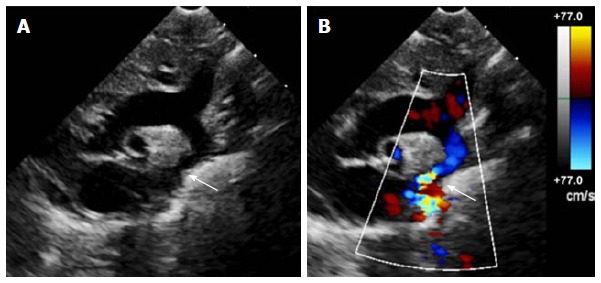
Figure 1.

Echocardiogram of coarctation. A: Two-dimensional transthoracic echocardiogram image obtained from the suprasternal notch in an 11-day-old infant demonstrating discrete coarctation (arrow); B: Color Doppler of the same image with aliasing of flow at the site of coarctation (arrow).
