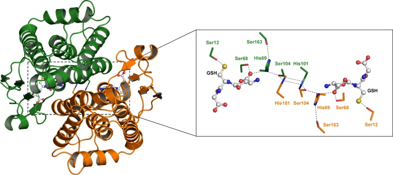
Figure 1. Ribbon representation of the D. melanogaster GSTE6 dimer.
Subunits (A) and (B) are distinguished by different colours, green and orange. The inset shows the ‘wafer’ histidine interface motif accompanied by serine residues connecting the two subunits from the active site of one subunit to the other. Ser12 is the catalytic residue.

