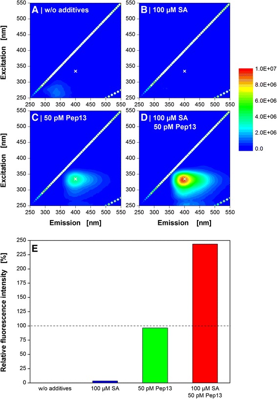
Fig. 2.

Fluorescence spectroscopy of Pep13-induced furanocoumarins in parsley cell suspension cultures. Cells were treated with the priming compound salicylic acid (SA) after 72 h and with Pep13 after 96 h. Cell culture supernatant was taken 24 h after the addition of Pep13 and subjected to 2D-fluorescence spectroscopy. Experimental conditions: quartz cuvette, 3 mL filling volume, spectral range of 250–550 nm. Reference cultivations: w/o additives (a), with the addition of exclusively 100 μM SA (b), and with the addition of exclusively 50 pM Pep13 (c). The priming experiment includes the addition of 100 μM SA and 50 pM Pep13 (d). All experiments are compared in (e) at an excitation wavelength of 335 nm and an emission wavelength of 400 nm (marked with a white cross in a-d). Reference cultivations: w/o additives (black column), with the addition of exclusively 100 μM salicylic acid (SA) (blue column), and with the addition of exclusively 50 pM Pep13 (red column). The total fluorescence signals from the supernatants of the cultures were subtracted by the total fluorescence signal of the untreated culture. Thereupon, the corrected fluorescence intensities of “100 μM SA” (blue column) and “50 pM Pep13” (green column) were set to 100 %. The priming experiment contained both the addition of 100 μM SA and 50 pM Pep13 (red column)
