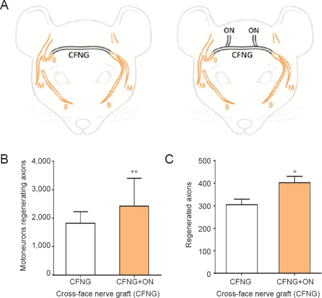
Figure 4.

Insertion of 2 sensory neurons in an end-to-side fashion into a denervated nerve stump improves the regeneration of axons through the nerve stump.
(A) Under halothane anesthesia and using aseptic technique, sensory occipital nerves were coapted end-to-side to the 30 mm long cross-face common peroneal nerve graft (CFNG) that connected the proximal stump of the transected right buccal branch of the facial nerve and the distal stumps of the transected left buccal and marginal mandibular branches. The motoneurons that regenerated their axons into the two left branches were backlabelled by exposing the two branches 10 mm distal to the CFNG to fluorogold and rubyred retrograde dyes for one hour, 16 weeks after the first surgery. (B) The number of motoneurons regenerating their axons across the CFNG was significantly increased after ‘protection’ of the chronically denervated distal facial nerve stump by the ingrowth of sensory neurons from the two sensory occipital nerves (ON) that were sutured to the side of the CFNG. (C) Regenerated axons enumerated from cross-sections of the buccal and marginal mandibular branches (taken ~11 mm distal to the CFNG), were also significantly increased in number when compared to the number of axons that regenerated through the unprotected distal stump of the facial nerve. The data are expressed as the mean ± SE, the stars representing a significant increase when sensory occipital nerves were coapted end-to-side (n = 12 for each of the CFNG and CFNG + ON groups; *P < 0.05; **P < 0.01).
