Figure 1.
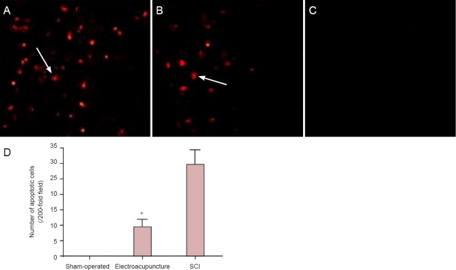
Effects of electroacupuncture on cellular apoptosis in injured rat spinal cord tissue.
(A–C) Apoptotic cells in the injured spinal cord tissue (TUNEL staining, × 200). Specific brown-yellow granules in the nuclei of cells were indicative of apoptosis. Apoptotic cells were present at the site of injury as well as in the surrounding regions. (A) Spinal cord injury (SCI) group; (B) electroacupuncture group; (C) sham-operated group. Arrows point to apoptotic cells. (D) Quantification of apoptotic cells in the injured spinal cord tissue. Data are expressed as the mean ± SD of five rats per group per time point. One-way analysis of variance was used for mean comparisons among groups, and two-sample t-test was used for mean comparisons between groups. *P < 0.05, vs. SCI group.

