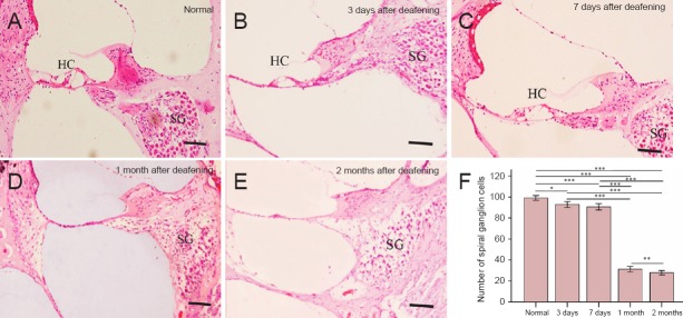
Figure 4.
Hematoxylin-eosin staining of frozen sections of the cochlea (A–E) and spiral ganglion (SG) cell counts (F) in untreated rats and in rats given injections of furosemide and kanamycin sulfate (Furosemide 200 mg/kg + Kanamycin 100 mg/kg group) at 3 and 7 days and 1 and 2 months after drug administration.
The morphology of cochleae in the control group (A) and in the Furosemide 200 mg/kg + Kanamycin 100 mg/kg group (B–E). Hair cells (HCs) had almost fully disappeared, but SG cells were intact at 3 and 7 days after drug administration. HCs were completely absent, and most SG cells were lost at 1 and 2 months after drug administration. Scale bars: 20 μm. (F) The SG cell count in the control and Furosemide 200 mg/kg + Kanamycin 100 mg/kg groups at 3 and 7 days and 1 and 2 months after drug administration. The data are expressed as the mean ± SD (n = 6). ***P < 0.001, **P < 0.01, *P < 0.05.

