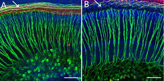
Figure 5.

Immunofluorescence staining of the whole cochlear basilar membrane preparation in the control group (A) and the Furosemide 200 mg/kg + Kanamycin 100 mg/kg group (B) 7 days after drug administration.
The number of auditory nerve fibers in the Furosemide 200 mg/kg + Kanamycin 100 mg/kg group was reduced compared with the control group. In the control group, a complete row of IHCs and three rows of OHCs were visible. The arrows point to the contacts between HCs and nerve fibers. After injury, the nerve fibers were damaged. Green fluorescence indicates the nerve fibers and SG cells stained with neurofilament-specific antibody, and the blue fluorescence indicates Hoechststained cell nuclei. Scale bars: 20 μm. IHC: Inner hair cell; OHC: outer hair cell; HC: hair cell; SG: spiral ganglion.
