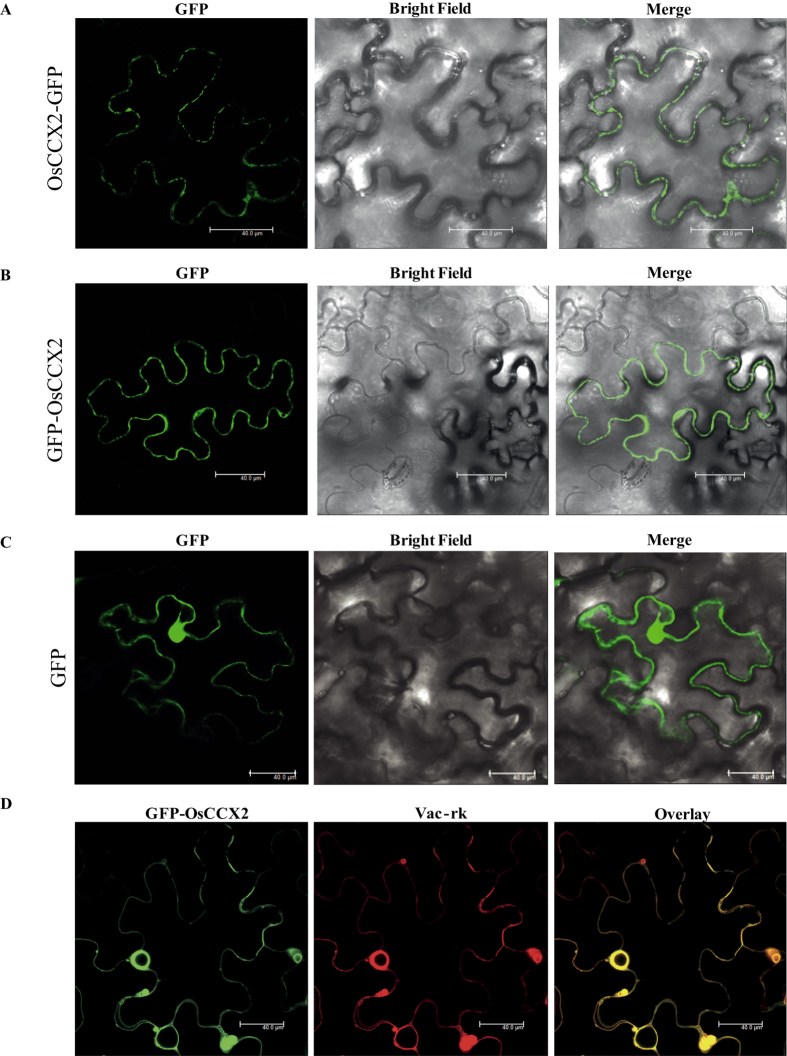
Figure 2. Subcellular localization of OsCCX2 proteins in Nicotiana benthamiana epidermal cells.
(A) OsCCX2 fused with N-terminal of GFP appears as circular pre-vesicles inside the lumen of the cell and seems to be localized to vacuolar membrane. (B) The OsCCX2 fused at the C-terminal of GFP also showed tonoplast localization. (C) Cells transformed with CaMV 35S -GFP were used as a control. Fluorescence was detected under a confocal laser-scanning microscope (wavelength: 488 nm). (D) The GFP-OsCCX2 co-localized completely with globular vesicles as shown by tonoplast markers (vac-rk). GFP fusions to OsCCX2 proteins are shown in green, mCherry vacuole markers are shown in red and overlay of two-mentioned proteins in dark field view. Scale bar = 40 μm.

