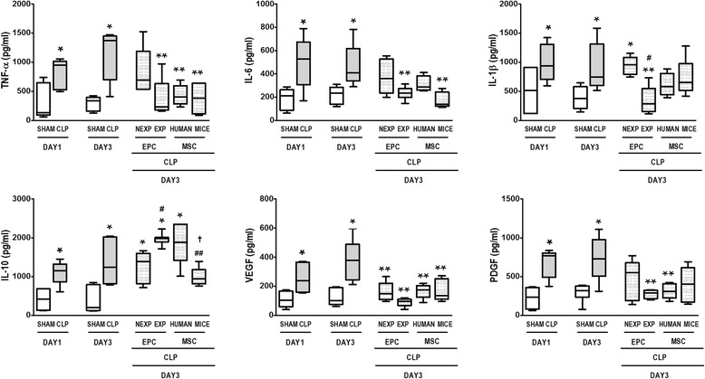
Fig. 3.

Lung inflammation on days 1 and 3. Lung tissue protein expressions of tumor necrosis factor (TNF)-α, interleukin (IL)-1β, IL-6, IL-10, vascular endothelial growth factor (VEGF), and platelet-derived growth factor (PDGF). Cecal ligation and puncture (CLP) animals were randomized to receive saline or non-expanded endothelial progenitor cells (EPC-NEXP), expanded endothelial progenitor cells (EPC-EXP), human mesenchymal stem cells (MSC-HUMAN), or mouse mesenchymal stem cells (MSC-MICE), intravenously. Values expressed as a box-and-whiskers plot of eight animals in each group. *P < 0.05, versus respective sham group. **P < 0.05, versus CLP group at day 3. # P < 0.05, versus EPC-NEXP group. ## P < 0.05, versus EPC-EXP group. † P < 0.05, versus MSC-HUMAN group
